Showing posts with label STPM. Show all posts

Monday, 19 April 2021

STPM Biology Biological Molecules Part 20 Osmotic, Turgor, Wall Pressure and Water Potential

Osmotic Pressure

  • When a solution is separated from pure water by semi-permeable membrane, there will be net water moving across into the solution.
  • The minimum pressure that has to be exerted by the solution to prevent water from moving in is called the osmotic pressure of the solution.

Osmotic pressure examples

Turgor Pressure

  • Turgor pressure = the pressure of cytoplasm exerted against the walls of a turgid cell.
  • This pressure is counteracted by the wall pressure.


Wall Pressure (Ψ p)

  • Wall pressure = the pressure of the cell wall exerted against the cytoplasm of the plant cell.
  • The wall pressure is also known as pressure potential (Ψ p) for plant cells.
  • Pressure potential usually has a positive value.

The relationship between turgor pressure and wall pressure

Water Potential (Ψ)

  • Water potential = the potential of water to move out of a solution by osmosis.
  • The water potential of a cell is the potential of water to move out of a cell through osmosis.
  • Symbol = (Ψ) ; Unit = kPa (kiloPascal; 1kPa = 1000Pa) or MPa (MegaPascal), 1MPa = 100,000Pa)
  • Pure water has the highest water potential. The water potential of pure water is 0 kPa at atmosphere pressure (101325 kPa).
  • The water potential of a plant cell (Ψ) = solute potential (Ψ s) + pressure potential (Ψ p)

The water movement from a dilute to a concentrated solution

Solute Potential (Ψ s)

  • Solute potential = the potential of a solution to take in water by osmosis due to the presence of solute materials.
  • Solute potential is also known as osmotic potential.


Water Potential for Solution (Ψ sol)

  • (Ψ sol) = the potential of water to move out of a solution by osmosis.
  • The water potential of a solution is negative in value.
  • This is because water potential for pure water is 0 kPa and pure water has the highest water potential.
  • Solution with larger negative water potential value have low water potential.
  • For example, cell A with water potential of -0.5 kPa has higher water potential than cell B with water potential value of -0.9 kPa.  Thus, water will flow from A to B.

The movement of water through different types of solution

STPM Biology Biological Molecules Part 19 Mineral Ions and Vitamins

  • Mineral ions and vitamins are generally needed in minute amounts. 
  • Lack of them in diet can lead to a variety of disorders.
  • The importance of mineral ions and vitamins are shown in the tables below.
Mineral ions
Mineral ions


Vitamins
Vitamins


Sunday, 11 April 2021

STPM Biology Biological Molecules Part 18 Nucleic Acids - DNA and RNA

DNA - the hereditary material of life

Structure

  1. A DNA molecule consists of 2 polynucleotide chains coiled to form a double helix. 
  2. The 2 chains are held together by hydrogen bonds between complementary bases. (T and A are complementary bases; while G and C are another complementary bases).
  3. Each complete turn of the double helix is 3.4nm long and contains 10 pairs of bases.
  4. The diameter of each helix is 1.0nm.
  5. The polynucleotide chains of DNA molecule are anti-parallel (the 5'end of one chain lies next to the 3' end of the other chain).
  6. the polynucleotide chain is made up of deoxyribonucleotides that are linked together by phosphodiester bonds.
Structure of DNA molecule

Complementary base pairing

  1. Due to its structure, only purine bases can pair with pyrimidine bases.
  2. Adenine (A, a purine) pairs with thymine (T, a pyrimidine) with 2 hydrogen bonds.
  3. Guanine (G, a purine) pairs with cytosine (C, a pyrimidine) with 3 hydrogen bonds.

Complementary base pairing


RNA

1. RNA is a polynucleotide. The monomer of RNA is ribonucleotide.

2. RNA molecule consists of one polynucleotide chain.

3. There are three types of RNA in cells:

  • ribosomal RNA (rRNA)
  • messenger RNA (mRNA)
  • transfer RNA (tRNA)


Ribosomal RNA (rRNA)

1. More than 80% of RNA in the cell is rRNA.

2. rRNA is found in ribosome. A ribosome is constructed from rRNA (50%) and protein (50%).

3. Functions of rRNA:

  • Main component of ribosome.
  • Bind mRNA molecule to ribosome during protein synthesis.


Messenger RNA (mRNA)

1. The longest RNA molecules which contains 70 to 3,000 nucleotides.

2. mRNA molecules are long uncoiled molecules.

3. Function of mRNA:

  • Carry genetic information from gene into the cytoplasm for protein synthesis.


Transfer RNA (tRNA)

1. The shortest RNA molecules.

2. tRNA makes up about 10% to 15% of RNA in cells.

3. The polynucleotide chain is folded to form a 'clover-leaf'.

4. The anticodon of tRNA contains 3 bases which are complementary to the codon for the amino acid it carries.

5. Function of tRNA:

  • Transfer a specific amino acid to ribosome for polypeptide synthesis.

rRNA tRNA mRNA
rRNA, mRNA, tRNA


Differences between RNA and DNA


Friday, 2 April 2021

STPM Biology Biological Molecules Part 17 Nucleic Acids

1. The 2 nucleic acids in the cells are:

  • DNA (deoxyribonucleic acid)
  • RNA (ribonucleic acid)

2. Nucleic acids are natural polymers. Nucleic acid monomers are nucleotides.

3. A nucleotide has 3 components:

  • 5-carbon sugar (pentose)
  • Organic base / nitrogenous base
  • Phosphoric acid

Structure of nucleotide


4. The pentose of nucleotides are ribose or deoxyribose. 

5. Nucleotide containing ribose is called ribonucleotides (RNA monomers).

6. Nucleotide containing deoxyribose is called deoxyribonucleotide (DNA monomers).

Structure of ribose and deoxyribose


7. Nucleotide has one of these five organic bases:

  • adenine (A)
  • guanine (G)
  • thymine (T)
  • cytosine (C)
  • urasil (U)

8. These bases can be divided into 2 group:

  • Purines (double-ringed molecule): Adenine, Guanine
  • Pyrimidine (single-ringed molecule): Thymine, Cytosine, Uracil

5 organic bases
5 organic bases


Formation of nucleotides and nucleic acid

  1. In formation of nucleotide, a nitrogenous base is first linked to pentose by condensation reaction to form nucleoside.
  2. Phosphate group is then added to the nucleoside to form a nucleotide.
  3. Nucleic acids are polynucleotides. Polynucleotides are formed by linking nucleotides together.
  4. Two nucleotides are linked together through condensation reaction to form dinucleotides.
  5. Further addition of nucleotides to dinucleotides will form polynucleotides.
  6. Nucleotides in polynucleotides are linked together in phosphodiester bonds.

    Formation of phosphodiester bond
    Formation of phosphodiester bond

  7. A nucleotide chain has 5' end and a 3' end. The 5' end of the polynucleotide chain is the end with the free phosphate group.

5' end and 3' end of nucleotide chain

Friday, 26 March 2021

STPM Biology Biological Molecules Part 16 Amino Acids - Properties of Protein

1. Protein is amphoteric.

  • Its structure has basic and acidic group.
  • Amino group, NH2 is basic; while carboxyl group, COOH is acidic.

Structure of amino acid

2. Protein is an important buffer in biological systems.

  • The amphoteric nature of protein allows it to function as a buffer.
  • Amino groups of protein removes excess acids in the system.
  • Carboxyl groups neutralize the excess bases in the system.

Protein as buffer in biological system

3. The colloidal nature of proteins allow it to exist as individual molecules in solution.

  • Colloids are particles not soluble in water but remain suspended in the solution.
  • Colloidal particles are usually 1nm to 100nm in diameter.
  • Most globular proteins are soluble in water due to the small size of its molecule and existing polar groups such as -COOH.
  • Globular protein with larger molecules will from colloidal suspensions in water.
  • The colloidal nature of protein provides a larger surface are for biochemical reactions in cells.

Differences between solution, colloidal solution and suspensions

4. Protein denaturation.

  • The change of structure and shape of the protein molecule due to the breaking down one or more bonds maintaining the structure of protein molecule is called protein denaturation.
  • It can be caused by acids, bases, heat, pH and ultraviolet light.

Protein denaturation

STPM Biology Biological Molecules Part 15 Amino Acids - Levels and Composition of Protein Structure

Levels of protein structure

1. Primary structure

  • Primary structure is structure showing the number and sequence of amino acids in its molecule.

Primary structure of insulin

2. Secondary structure

  • Secondary structure is the structure showing the coiling of polypeptides to become helix or the folding of polypeptide to become pleated-sheet.
  • Secondary structures are maintained and stabilized by hydrogen bonds.

Secondary structure of protein

3. Tertiary structure

  • Tertiary structure of protein is the structures showing how a single polypeptide chain (helix) is folded form a globular structure.
  • This structure is maintained by various bonds; among which are disulphide bonds, electrovalent bonds or hydrogen bonds.

Tertiary structure of protein

4. Quaternary structure

  • Quaternary structures of protein is the structure showing how two or more polypeptide chains are bound together.
  • Examples of protein with quaternary structure are hemoglobin.


Quaternary structure of protein


Four levels of protein structure


Composition and structures of protein

1. Proteins can be classified according to its composition or structure.

2. Based on composition, proteins can be grouped into:

  • simple protein
  • conjugated protein

3. Based on structure, proteins can be divided into:

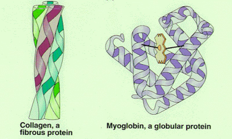
  • Fibrous protein
  • globular protein


Simple protein

  • Proteins that contain amino acids only.
  • For examples: albumin, globulin, and histone.


Conjugated protein

  • Protein bounds to non-protein groups.
  • Non-protein groups which bound to proteins are known as prosthetic groups.
  • For example: hemoglobin.

Structure of hemoglobin
Structure of hemoglobin

Fibrous protein

(a) Fibrous protein consists of long and parallel polypeptide chains.

(b) The polypeptide chain in fibrous protein is usually coiled to form α-helix.

(c) Neighboring helical chains are usually cross-linked by hydrogen bonds, electrovalent bonds, or disulphide bonds.

(d) Fibrous protein are not soluble in water and are very strong.

(e) Examples of fibrous protein:

  • Collagen - in tendons, cartilages, bones and skin.
  • Myosin - structural protein in muscles.
  • Keratin - structural protein in hairs, nails, feathers and horns.
  • Elastin - found in ligaments.
  • Sclerotin - combines with chitin to form the exoskeleton of insects.

(f) Collagen is the most common protein in mammals.

  • This protein is a main structural component of connective tissues (cartilage, skin, tendons and ligaments).
  • The basic structure of collagen is a tropocollagen helix which consists of three polypeptide chains (helixes) twisted together.
  • The chains are stabilized by hydrogen bonds between the protein chains.

Collagen fiber
Collagen fiber


Globular protein

(a) In globular proteins, the polypeptides (helixes) are folded into globular structures.

(b) The globular structure is maintained and stabilized by hydrogen bonds, disulphide bonds and electrovalent bonds.

(c) Some globular proteins are soluble in water; some of them form suspension, and the rest are insoluble in water.

(d) Globular proteins are easily denatured. This is because the hydrogen bonds and disulphide bond in the molecule can easily be broken.

Denaturation of protein


(e) Examples of globular protein:

  • Hemoglobin
  • Myoglobin
  • Hormones
  • Enzymes

Tuesday, 16 March 2021

STPM Biology Biological Molecules Part 14 Amino Acids - Structure and Functions of Protein

1. Two amino acids can linked together to form a dipeptide by a condensation reaction. In the dipeptide, the two amino acids are linked by a peptide bond.


Formation of peptide bond


2. Peptide bond can only be formed by condensation reaction between the carboxyl group of one and the amino group of another amino acid.
3. Further amino acids can be added on either end of the dipeptide to form a polypeptide. Proteins consists of one or more polypeptides.
Polypeptide chain
Polypeptide chain


Structure of protein
  1. A protein may contain up to 20 types of amino acids. 
  2. Amino acids in proteins are linked together by peptide bond.
  3. Proteins are polymers with a large relative molecular mass.
  4. Protein molecules may contain one or more polypeptide chains.
  5. A polypeptide chain has a carboxyl group, COOH at one end and an amino group, NH2 at the other end (refer to the diagram above).

Fibrous proteins
(a) In fibrous proteins, the polypeptide chains exist as long parallel helixes. Helixes are cross-linked by:
  • hydrogen bonds,
  • disulphide bonds, or
  • electrovalent bonds
(b) Examples of fibrous proteins are collagen, keratin and fibrin.


Globular proteins
(a) In globular protein, the polypeptide chains (helix) are tightly folded to form a globular molecules. 
(b) The globular structure is stabilized by cross links, such as disulphide bonds.
(c) Examples of globular proteins include enzymes, myoglobin, insulin and hemoglobin.
fibrous protein globular protein

Example of fibrous and globular protein


Conjugated proteins
(a) A protein may conjugate with prosthetic groups such as lipid, polysaccharides, nucleic acid, metals and ions.
(b) Examples of conjugated proteins are lipoprotein, nucleoprotein, glycoprotein and hemoglobin.

Conjugated protein
Conjugated protein


Functions of protein

  1. Protein is an important structural construction material.
  2. Protein can also function as catalyst. Enzymes are proteins that catalyze biochemical reactions.
  3. Transport of oxygen and carbon dioxide. Transport proteins include hemoglobin, the carrier of oxygen in blood.
  4. Maintain pH of cytoplasm and blood.
  5. Proteins are stored in organisms for food and energy.
  6. Protein is also involved in the defense and protection of body against pathogens. Antibodies are proteins in the blood that destroys pathogens. Fibrinogen and prothrombin are involved in blood clotting. Mucus protects the epithelial layer of digestive organs.
  7. Functions as hormones. Insulin, prolactin, and thyroxin are all proteins.
  8. Protein is also involved in food digestion. Digestive enzymes are proteins.
  9. Proteins play an important role in movements and mobility. Actin and myosin are involved in muscle contraction and movements of an amoeba. 
  10. Protein such as thyroxine is important for growth.

Wednesday, 10 March 2021

STPM Biology Biological Molecules Part 13 Amino Acids

1. Protein is an organic compound consisting of carbon, hydrogen, oxygen, nitrogen and sometimes sulphur and phosphorus.

2. Protein is a natural polymer, the monomer of protein is amino acid.

3. The basic structure of amino acid:

Basic structure of amino acid

4. Amino acid has a basic amino group, -NH2 and a acidic carboxyl group, -COOH and a side chain, R. There are 20 types of amino acids found in proteins. Different amino acids have different R chain.

Amino Acids
Three Letter Abbreviation
Alanine
Ala
Arginine
Arg
Asparagine
Asn
Aspartic acid
Asp
Cysteine
Cys
Glutamine
Gln
Glutamic acid
Glu
Glycine
Gly
Histidine
His
Isoleucine
Ile
Leucine
Leu
Lysine
Lys
Methionine
Met
Phenylalanine
Phe
Proline
Pro
Serine
Ser
Threonine
Thr
Tryptophan
Trp
Tyrosine
Tyr
Valine
Val


5. Amino acids can be classified into 4 major types based on the side chain (R).

  • Non-polar amino acids
  • Polar amino acids
  • Basic amino acids
  • Acidic amino acids


Non-polar amino acids

  • Non-polar amino acids are amino acids with a non-polar side chain
  • Examples: glycine, alanine, valine, leucine, methionine, isoleucine, phenylalanine, and tryptophan.
Non-polar amino acids


Polar amino acids

  • Polar amino acids are amino acids with a polar side group.
  • Examples: serine, threonine, asparagine, glutamine, tyrosine, and cysteine.
    Polar amino acids
    Polar amino acids


Basic amino acids

  • Basic amino acids have side chain which is an amino group, NH2, a base group. 
  • Examples: lysine, arginine, and histidine.

Basic amino acids
Basic amino acids


Acidic amino acids

  • Acidic amino acids have a side chain which is a carboxylic group, COOH.
  • Examples: aspartic acid and glutamic acid.
Acidic amino acids
Acidic amino acids

Sunday, 7 March 2021

STPM Biology Biological Molecules Part 12 Lipid - Steroids

1. Steroid is very different in structure compared to lipid but is classified together with lipid because of their similar nature.

2. Steroid are abundant in animal tissues, but it is hardly found in plants.

3. All steroid molecules consist of four fused rings of carbon atoms. Different steroids have different functional groups attached to this ensemble of rings.


The typical steroid structure with 17 carbon atoms


4. The most common steroid in the body is cholesterol. Many steroid hormones are produced from cholesterol.

5. Other examples are vitamin D2 (calcipherol), bile acids, sex hormones (testosterone, progesterone and oestrogen), and adrenaline.

Examples of steroids

6. Cholesterol is synthesized in the liver. The importance of cholesterol in health:

  • Cholesterol is important in the health of cell membrane. It is a major component in the cell membranes of animals.
  • Cholesterol is a precursor for synthesizing animal hormones such as progesterone, bile acids, oestrogen, and others. These hormones are important for the healthy functioning of organisms.
  • Used to synthesize vitamin D in the skin.
  • Excess cholesterol in the body can lead to arteriosclerosis, high blood pressure and heart attacks.

Arteriosclerosis
Arteriosclerosis

STPM Biology Biological Molecules Part 11 Lipid - Phospholipids

1. Phospholipid molecule is formed from the condensation of:

  • one glycerol molecule
  • two fatty acids molecules
  • one phosphoric acid molecule


Structure and symbol of a phospholipid molecule


2. Phospholipid is a major component of cell membrane and hence is distributed all over the body of an organism.


Structure of phospholipid molecule

  1. A phospholipid molecule consists of one glycerol molecule attached to two fatty acids chain and one phosphate group.
  2. Additional small molecules (usually polar) can be linked to the phosphate group to form a variety of different phospholipids.
  3. For instance, choline molecule attaches to the phosphate group of phospholipids to form lecithin. 
  4. Lecithin is the most common phospholipid in human bodies. The major phospholipids of cell membrane is lecithin. The fatty acids of lecithin is oleic acid (unsaturated) and stearic acid (saturated).

Structure of lecithin


Physical properties of phospholipid

  1. The hydrophilic head of phospholipid molecules is soluble in water, while the hydrophobic tail is insoluble in water.
  2. In water, phospholipid molecules assemble into a droplet called micelle. On the surface of water, phospholipid molecules are assemble closely with hydrophilic heads dissolved in water and the tails jutting out of the surface.
    Assembly of phospholipid molecules on the water surface and in water
    Assembly of phospholipid molecules on the water surface and in water

  3. In cells, both the intracellular environment and the immediate external environment are water. This causes phospholipids to form bilayer. In this bilayer, the hydrophilic heads are on the outside (in contact with water); the tails of phospholipids point to the interior. This bilayer is the main structures in the formation of cell membrane.
    Cross section of a phospholipids bilayer
    Cross section of a phospholipids bilayer


Functions of phospholipid

  1. Phospholipids are the main structural components for cell membranes.
  2. Phospholipid such as lecithin is involved in the production of neurotransmitters such as acetylcholine in the neurons.
  3. Phospholipid molecules are usually involved in transporting fat in the body.

Monday, 1 March 2021

STPM Biology Biological Molecules Part 10 Lipid - Triglycerides

1. All fats and oils are triglycerides.

2. A triglyceride molecule is formed through condensation reaction between three fatty acids and one glycerol molecule.

3. Hydrolysis of a triglyceride molecule produces fatty acid molecules and glycerol molecule.

Condensation and hydrolysis reactions of triglyceride


4. Each fatty acid molecule is linked to glycerol by an ester bond, -COO. 

Ester bond, -COO
Ester bond, -COO


5. Saturated fat is a fat that does not contain any C=C bond in its molecule. Most of the saturated fat exists as solids in room temperature.

6. Unsaturated fat is a fat that has one or more C=C bonds in its molecule.

  • Monounsaturated fat is fat with molecules that only has one double bond between its carbon atoms. For examples, sesame oil and groundnut oil.
  • Polyunsaturated fat is fat with molecules that have two or more doubles C=C bonds in its molecules. For examples, maize oil, linseed oil and cottonseed oil.
  • Polyunsaturated fat has a low boiling level, thus it exists as liquid in room temperature.

Saturated, monounsaturated and polyunsaturated fat molecules


7. The consumption of food rich with saturated fat and cholesterol increases the risks of cardiovascular diseases. Saturated fat molecules and excess cholesterol deposit on the artery walls, thus narrowing it.


Physical properties of triglycerides

  • Not soluble in water, but soluble in organic solvents.
  • High relative molecular mass.
  • High ratio of hydrogen atoms to oxygen atoms in molecule.
  • Can be emulsified.
  • Fat usually exists as a solid while oil exists as liquid at room temperature.


Chemical properties of triglycerides

  • Triglycerides undergo hydrolysis to form fatty acids and glycerol.


Distribution of triglycerides in organisms

  • In adipose tissues underneath the skin.
  • Attached to the surface of visceral organs such as the heart, liver and digestive tracts.
  • In eggs.
  • In seeds (maize, peanuts and etc.).


Functions of triglycerides

  • Source of energy of organisms.
  • As food and energy stores for animals.
  • Major components of plasma membrane.
  • Heat insulation. Fat underneath the skin is a heat insulator for animals in cold region.
  • Water proofing. Oils and waxes on the outer surface of organisms waterproof the body.
  • Protection. Fats packed around visceral organs protect these organs.
  • Fat is a source of metabolic water for animals.
  • Fat is an important solvent for vitamins A,D,E,K and hormones in the body.
  • Saturated fat is the raw material for synthesizing cholesterol.
  • Buoyancy. Stored fats also aid buoyancy.